


With spinal column osteochondrosis, many are familiar with popular shows from the TV screen but from their own unfortunate experience.Statistics are strict: up to 80% of the population suffer from this disease, which is also significantly young.If the previously complained of spine problems was mainly among the older generation, osteochondrosis of children is now no surprise.And sedentary lifestyle fault and so written "the benefits of civilization".

Osteochondrosis of the cervical spine is a polyethological progressive disease, which is manifested by the degeneration of the intertectebral discs of the spine.Everyone knows about the symptoms, but this knowledge is fragmented;We will try their structure as well as talk about the principles of diagnosis and treatment of osteochondrosis of the cervical spine.
Causes of osteochondrosis
Medical science cannot be answered unequivocally, which is why osteochondrosis.It is reliably known that the seductive lifestyle that modern man is prone to adversely affect the progression of this disease.Interestingly, both hypodynamics and the colossal loading of athletes lead to a puppet of the discs.The hereditary factor plays a major role.The following reasons are distinguished:
- Hereditary history;
- Obesity;
- Hypodynamia;
- Metabolic disorders in the body;
- Traumatic spinal column injury;
- Long static overload and work associated with weight gain (working on computer, weight gain, miners, movements, etc.);
- Scoliosis;
- Dysfunctional environmental condition;
- Flat legs and pregnancy;
- Hypothermia and stress, which often leads to exacerbation of the disease.
There are several neurological syndromes:
- Shoulder -chedded parantharthritis;
- Root;
- Heart;
- Vail artery syndrome.
Shoulder -cheeky peritric.It is characterized by pain in the neck, shoulder, shoulder joint.The leading neurogenic contracture of the shoulder joint arises, which is protective in nature as it protects against the stretching of the axillary nerve (antalgic posture).In this position, the muscles surrounding the joint are in tension.The severity of pain syndrome depends on the degree of exacerbation of osteochondrosis: On the slight restriction of the amplitude of the joint movements on the so -called "frozen shoulder", when any movement causes severe pain.The pain becomes intense when the shoulder is diverted and expressed, as these movements exacerbate the tension of the axillary nerve.
Royshift syndrome (cervical sciatica).The most common is cervical osteochondrosis.At the same time, the spinal nerve spine is contracted due to the "subordination" of the intervertebral discs, as well as due to osteophyte growth or disk leakage in the lateral direction.Pain syndrome is specific: intense burning, tearing, pressing pain, which also becomes intense when the patient moves to the head.Antalgic posture is also observed in the neck muscles, they are sharply tense and painful, the volume of movements is limited.There is pain in the head, neck, front chest, shoulder, shoulder blades.Sensitivity is characterized by the type of "short jacket sleeve" type.
Cardial syndrome.The name of the syndrome itself is responsible: the clinical picture is very similar to the pectities of angina.In this case, there is no organic heart injury, at the height of pain syndrome, the ECG is not detected by coronary blood flow and such patients tolerate well.A typical feature of angina pectoror: Pain occurs after nitrate is administered, and in the case of osteochondrosis it does not change and suffer for a long time.Unlike angina, pain localization is mainly in the left heart.With irritation of the segments C8 - T1 roots, rhythm disorders are possible in the form of tachycardia and extrasystole.It is not caused by damage to the cardiac conduction system, but rather by disruption of sympathetic innervation of the heart muscle (extracardial injury).During the differential diagnosis of angina pectoror and heart syndrome, the lead is the fact that in addition to cardiac complaints, the patient indicates an increase in pain in the shoulder joint and neck associated with rough or coarse movements.
Vail artery syndrome.The spine artery occurs in the canal formed by the holes in the hole in the transverse processes of the spine.This artery is paired, it is responsible for blood supply to the brain.Consequently, any narrowing of this channel has a very negative effect on brain tissue nutrition.Spine artery syndrome develops directly both by the compression of the artery and with the irritation of the sympathetic nerve plexus, which is located around it.In this pathology, the pain is burning or pulsating in the back of the back, with the spread of whiskey, tutorial arcs, crows.It arises on both sides and on both sides.Patients usually associate exacerbation with a condition after sleep in a non -asiological posture, travel in transport, walk.With pronounced symptoms, hearing loss, dizziness, noise in the ears, nausea, vomiting, loss of consciousness and increased blood pressure.Such symptoms are not specific and are very similar to claims during cerebral stroke.This pathology is characterized by Sistine Chapel syndrome: reduced, which occurs when the head is retreated (a strong ischemia of the brain).He was described by visitors to Sistine Chapel in the Vatican when they examined the frescoes in the arches.It is also possible to fall without loss of consciousness, with sharp turns of the head.
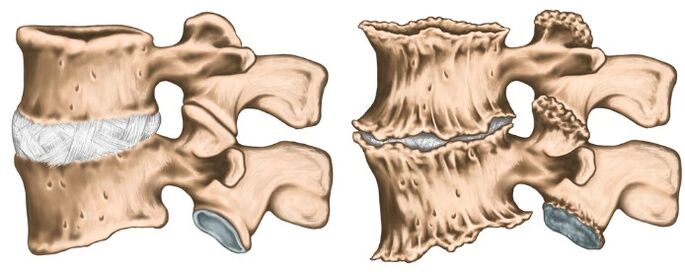
Like any diagnosis in medicine, the diagnosis of osteochondrosis has been established on the basis of patient complaints, disease anamas, clinical examination, and auxiliary research methods.The cervical spine X -ray is direct and lateral forecasts, if necessary, in special positions (open mouth).At the same time, experts are interested in the height of the intervertebral discs, the presence of osteophytes.Modern research methods use IAMR and CT examinations, which will allow the most accurate verification of the diagnosis.In addition to the following methods of additional studies, consulting specialists (cardiologist, ophthalmologist, neurosurgeon) may be required, and the neurologist's examination is simply vital.The neurologist is busy treating osteochondrosis, so after examining the patient, it determines the necessary minimal examination at its discretion.

Treatment of osteochondrosis
Osteochondrosis is a polyetic disease, one course of therapy has not been cured.You can't drink "magic pills" and everything will pass, it is necessary to fundamentally change your lifestyle, as the trigger is hypodynamic.The most tangible results at the initial stage of the disease are easier when complaints are minimal and there are no compression syndromes and spinal cord.At the acute stage of the disease, when the following groups of drugs are prescribed with pronounced pain: Pain syndrome is expressed:
- Therapeutic paravertebral blockade (to relieve pain and to remove muscle spasms);
- Nsaids;
- NSAIDs containing ointments and reflexive action;
- Muscle calmers;
- B vitamins V.
The inflammatory process stops and alleviates pain syndrome, they move on to the treatment of physio-therapy.Most often, the following techniques are used:
- Laser therapy;
- Electrophoresis;
- Acupuncture;
- Exercise therapy;
- Therapeutic massage;
- Manual therapy.
It is important to understand that osteochondrosis occurs with periods of exacerbation and remission, so it is very important to influence the cause, not the treatment of the investigation.